If a gray, dim, or distorted spot has appeared in the center of your vision in one eye, central serous chorioretinopathy — often abbreviated CSC or CSCR, and frequently misspelled "central serous choriotinopathy" in searches — is one of the most common explanations in otherwise healthy adults between 30 and 55. It is also one of the most misunderstood: usually self-resolving, yet sometimes chronic and vision-threatening, and strongly linked to medications many patients don't realize contain steroids.
What Is Central Serous Chorioretinopathy?
Beneath the retina lies the choroid, a dense layer of blood vessels that nourishes your central vision. In CSC, the choroid becomes thickened and hyperpermeable, and fluid pushes through the retinal pigment epithelium — the retina's waterproofing layer — collecting in a blister under the macula. The macula is the part of the retina responsible for reading, faces, and fine detail, which is why even a small pocket of fluid is so noticeable.
Symptoms Patients Actually Describe
- A round gray or brown smudge in the center of vision in one eye
- Metamorphopsia — straight lines (door frames, text lines, tile grout) look bent or wavy
- Micropsia — objects appear slightly smaller with the affected eye
- Colors looking washed out or dimmer on one side
- A delay in vision "catching up" after bright light
- Blurred vision that glasses do not fix — though a temporary mild farsighted shift is common
Symptoms usually develop over days. CSC is typically painless, and the eye looks completely normal from the outside.
Who Gets CSC — and the Steroid Connection
The classic patient is a man in his 30s to 50s under significant stress, but women and older adults get CSC too. The most important modifiable risk factor is corticosteroid exposure in any form:
- Oral prednisone or dose packs
- Joint, epidural, or intramuscular steroid injections
- Steroid inhalers for asthma or COPD
- Nasal steroid sprays for allergies
- Potent steroid skin creams over large areas
Other associations include high psychological stress, "type A" behavior patterns, Cushing syndrome, pregnancy, and sleep apnea. Part of every CSC evaluation is a careful medication review — patients are often surprised that an allergy spray or dermatologic cream is relevant to their retina.
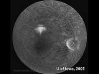
How We Diagnose It
CSC is confirmed quickly and painlessly:
- Dilated retinal examination — a shallow, well-demarcated dome of fluid at the macula
- OCT imaging — a cross-sectional scan that shows the subretinal fluid directly, measures it, and tracks it to resolution at follow-up visits
- Amsler grid testing — documenting the distortion you experience, and giving you a home tool to monitor change
When the picture is atypical — or treatment is planned — we coordinate fluorescein angiography or fundus autofluorescence with a retina specialist to map the leak. The key diagnostic responsibility is distinguishing CSC from conditions it can mimic, including wet macular degeneration in older patients, where the same fluid has a very different cause and urgency.
Treatment: When to Watch, When to Act
Acute CSC — structured observation
Most first episodes resolve on their own within 3 to 6 months, with good visual recovery. Management is active, not passive: stop or substitute every corticosteroid source where the prescribing physician agrees it is safe, address stress and sleep apnea, and re-scan with OCT until the fluid is gone.
Chronic or recurrent CSC — treatment
Fluid persisting beyond roughly 3–6 months, repeated recurrences, or early degenerative retinal changes shift the calculus, because long-standing fluid permanently damages the photoreceptors. The best-supported options, delivered through our retina specialist network:
- Half-dose photodynamic therapy (PDT) — targets the leaking, thickened choroid itself; the leading evidence-based treatment for chronic CSC
- Subthreshold micropulse laser — stimulates the retinal pigment epithelium to pump fluid out without burning the retina
We manage the diagnosis, steroid audit, monitoring, and co-management, and refer for PDT or laser at the right moment — so treatment happens neither too early (unnecessary) nor too late (irreversible).
What to Expect at Your Evaluation
A CSC workup at our office includes dilation, OCT imaging of both eyes (the fellow eye shows choroidal changes more often than patients expect), refraction to document any hyperopic shift, a complete steroid and medication review, and a written monitoring plan. Most visits take about an hour.
When to Seek Care Promptly
- Any new gray spot, distortion, or size difference in central vision
- Known CSC with vision worsening instead of stabilizing
- A new central blur if you are over 55 — wet macular degeneration must be ruled out quickly
- Central symptoms while using any steroid medication
Next Steps
If you're searching for a central serous chorioretinopathy evaluation near you in Orange County, Dr. Alexander Bonakdar, O.D., has diagnosed and co-managed retinal conditions in Santa Ana since 1991, with in-office OCT imaging and an established retina specialist network for the cases that need PDT or laser. Call (949) 323-3600 or book online — same-week appointments are available, and new central vision changes are prioritized.
Educational content — not a substitute for an individual exam. Never stop a prescribed steroid without talking to the prescribing physician; we coordinate that conversation as part of your care. Questions? Contact us.
Have Questions About Your Eye Health?
Dr. Alexander Bonakdar and his team are here to help. Schedule a consultation to discuss your specific needs.